
BARIUM X-RAY
This is for informational purposes only. For medical advice or diagnosis, consult a professional.
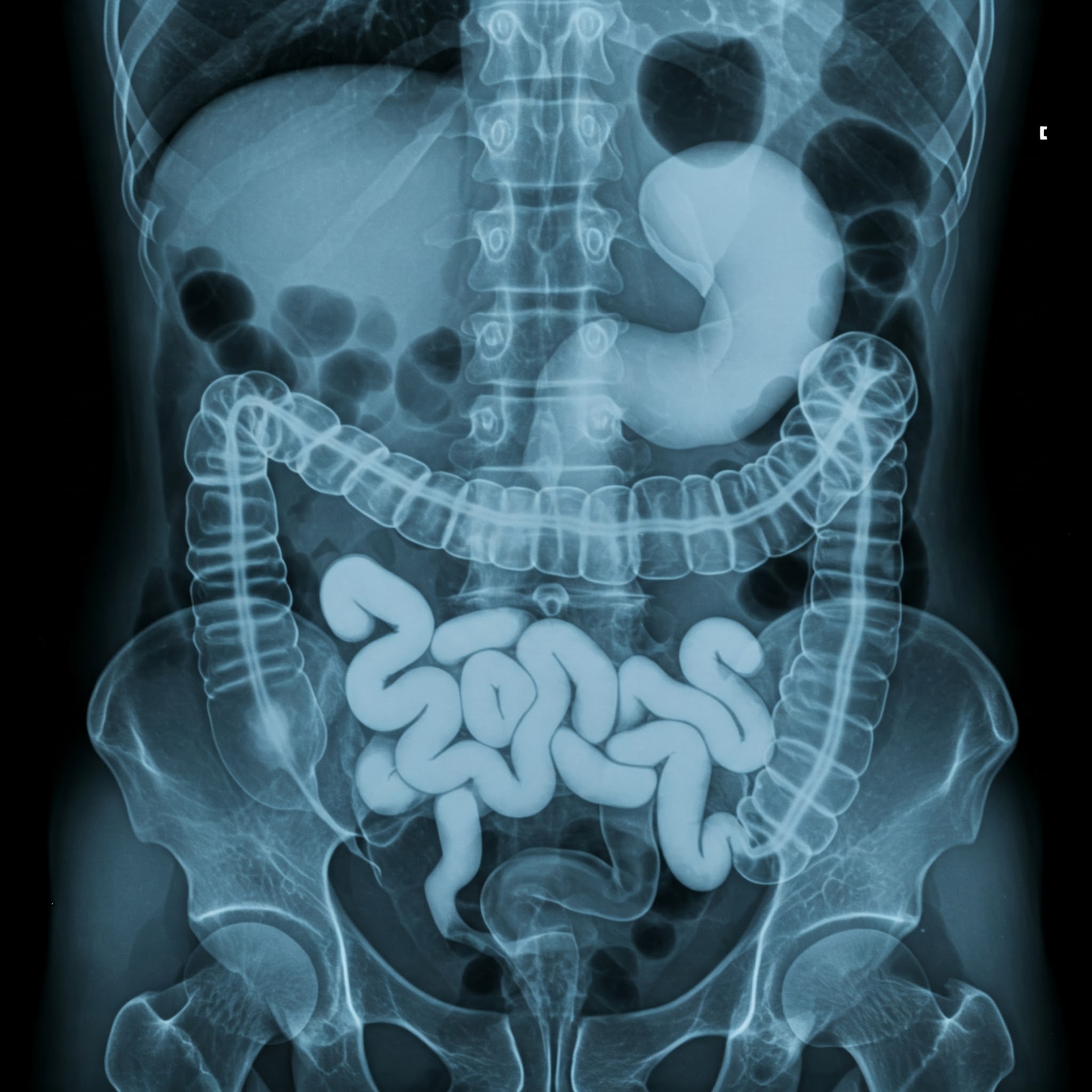
A barium X-ray is a type of imaging test that uses a special contrast material called barium to highlight the gastrointestinal (GI) tract, making it visible on X-ray images. Barium is a metallic chemical that appears white on X-rays and is used to visualize the:
Esophagus
Stomach
Small intestine
Large intestine (colon)
Types of Barium X-ray Procedures:
There are different types of barium X-ray procedures, depending on which part of the GI tract needs to be examined:
Barium swallow: Used to examine the esophagus. You will swallow a liquid containing barium.
Barium meal: Used to examine the esophagus, stomach, and first part of the small intestine (duodenum). You will drink a liquid containing barium.
Small bowel follow-through: Used to examine the small intestine. X-ray images are taken over time as the barium moves through the small intestine.
Barium enema: Used to examine the large intestine (colon). Liquid containing barium is inserted into the rectum through a tube.
Purpose:
Barium X-rays can help diagnose various conditions affecting the GI tract, such as:
Ulcers
Polyps
Tumors
Inflammatory bowel disease (Crohn's disease, ulcerative colitis)
Swallowing difficulties
Blockages
How it Works:
During the procedure, X-ray images are taken as the barium moves through your GI tract. The barium coats the lining of the GI tract, providing a clear outline of these organs on the X-ray images.
Risks:
Barium X-rays involve a small amount of radiation exposure. The risk associated with this exposure is generally considered low. However, it's important to inform your doctor if you are pregnant or think you may be pregnant, as radiation can pose a risk to the developing fetus.
Overall, a barium X-ray is a valuable diagnostic tool for evaluating the GI tract and detecting various abnormalities.
As low as
4000.00 2000.00


.jpg)


