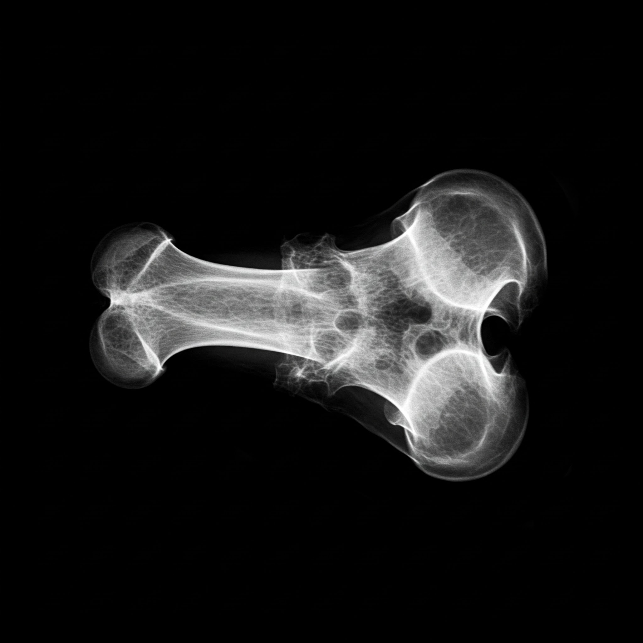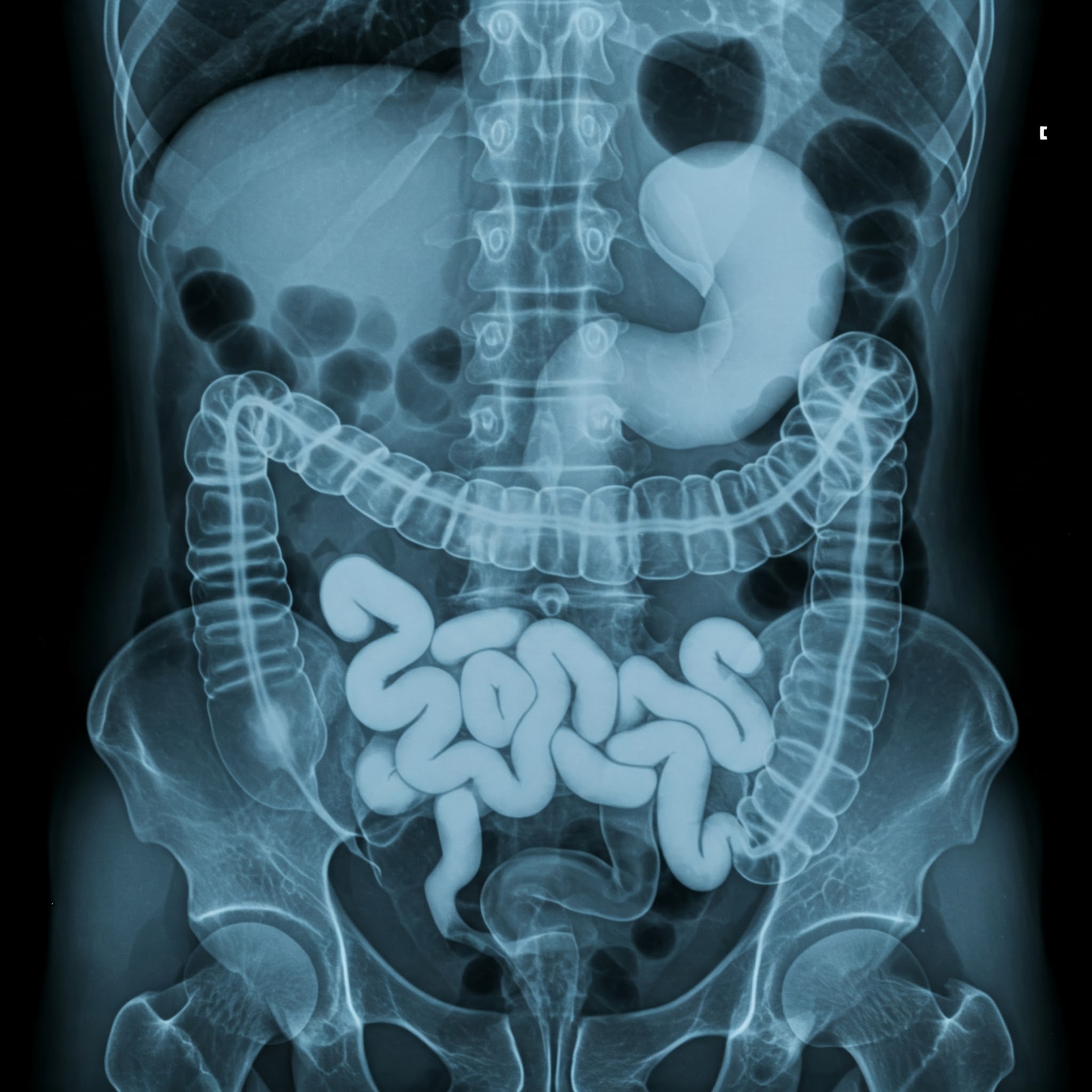

BONE X-RAY
This is for informational purposes only. For medical advice or diagnosis, consult a professional.
A bone X-ray is a quick and painless imaging test that uses small amounts of radiation to create images of your bones. It's a common and effective way for doctors to visualize and assess the condition of your bones.
How it Works
During a bone X-ray, you'll be positioned on a table or against a vertical surface, and the X-ray machine will send beams of radiation through the specific area of your body being examined. Because bones are dense, they block the passage of the X-ray beams, creating clear images on a detector. These images are then displayed on a computer screen for a radiologist to interpret.
What it Shows
Bone X-rays can reveal a variety of conditions and abnormalities, including:
Fractures: Bone X-rays are excellent at detecting breaks, cracks, and other types of bone fractures.
Dislocations: They can show if a bone has been displaced from its normal position in a joint.
Arthritis: X-rays can reveal signs of osteoarthritis, rheumatoid arthritis, and other forms of arthritis, such as joint space narrowing, bone spurs, and changes in bone density.
Infections: Bone infections (osteomyelitis) can cause changes in bone structure that are visible on X-rays.
Tumors: Both benign and malignant bone tumors can be detected with X-rays.
Bone loss: X-rays can help assess bone density and identify areas of bone loss, which can be a sign of osteoporosis.
Congenital abnormalities: Bone X-rays can be used to identify birth defects affecting the skeletal system.
As low as
500.00 250.00


.jpg)


