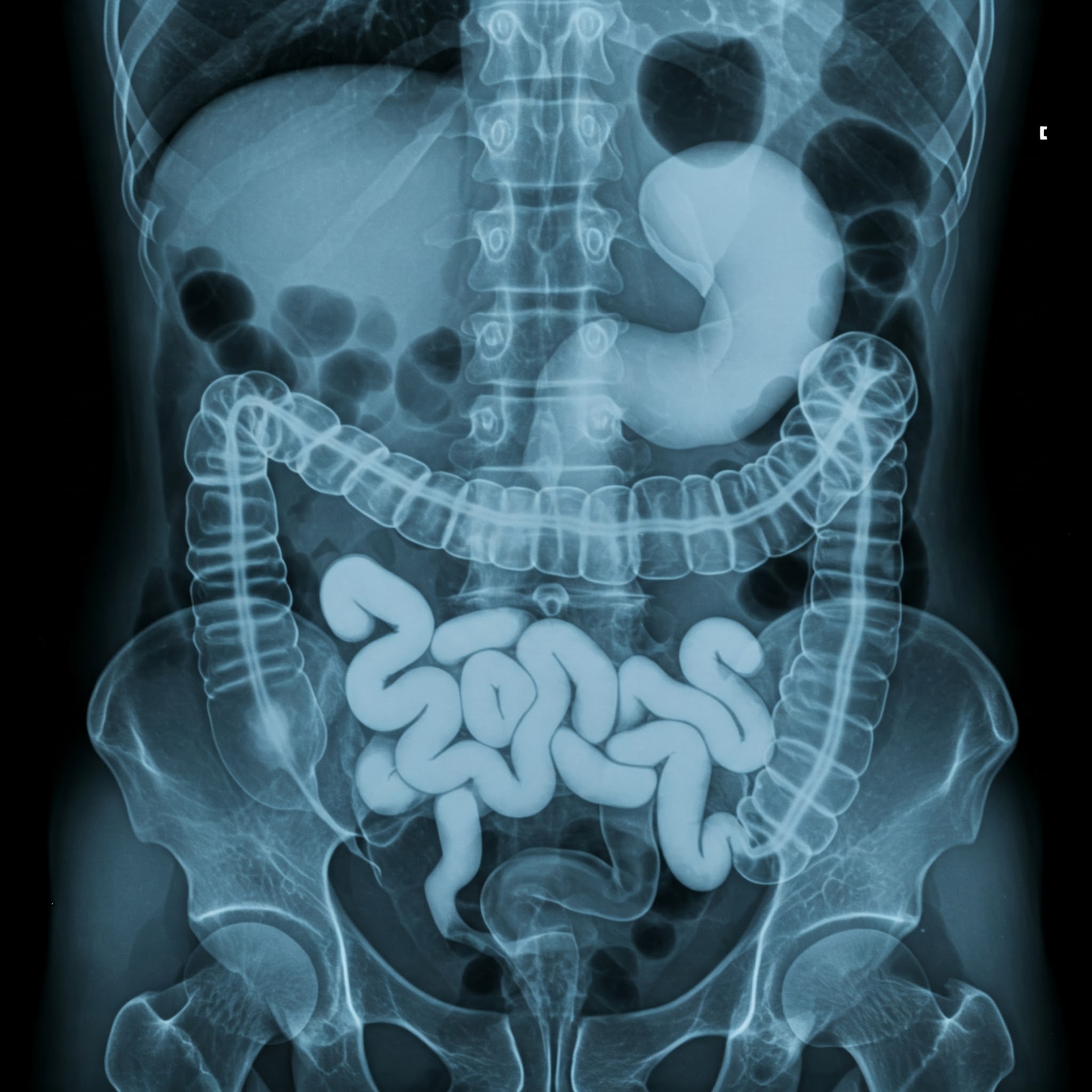

CHEST X-RAY
This is for informational purposes only. For medical advice or diagnosis, consult a professional.
A chest X-ray is a common imaging test that uses small amounts of radiation to create images of the structures in your chest, including your:
Lungs
Heart
Blood vessels
Airways
Bones of the chest and spine
How it Works
During a chest X-ray, you'll stand or sit in front of an X-ray machine. The technician will position you and may ask you to hold your breath for a few seconds while the images are taken. The X-ray machine sends radiation beams through your chest, and the images are captured on a detector. Dense structures like bones appear white on the image, while air-filled spaces like the lungs appear black.
What it Shows
Chest X-rays can help diagnose a wide range of conditions, including:
Lung infections: Pneumonia, bronchitis, and tuberculosis
Lung conditions: Lung cancer, chronic obstructive pulmonary disease (COPD), and collapsed lung
Heart problems: Heart enlargement and heart failure
Injuries: Fractures of the ribs or spine
Other conditions: Fluid around the lungs, air in the chest cavity, and abnormalities in the blood vessels
As low as
400.00 100.00


.jpg)


